
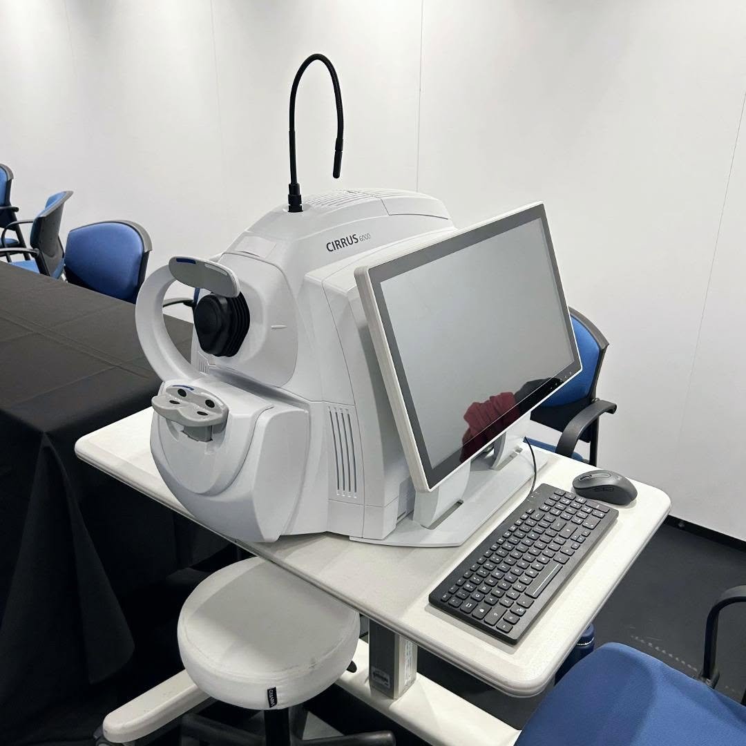
Product details
ZEISS CIRRUS 6000 Optical Coherence Tomography
The ZEISS CIRRUS 6000 Optical Coherence Tomography (OCT) is a high-performance OCT system designed for modern eye care practices that require fast imaging, detailed visualization, and efficient clinical workflows. With high-speed image acquisition, enhanced imaging detail, and a wider scanning field, CIRRUS 6000 supports clinicians in evaluating retinal and optic nerve structures with greater efficiency.
Designed for practices with demanding patient volumes, the CIRRUS 6000 combines high-speed OCT and OCT Angiography (OCTA) capabilities with advanced analysis tools to support clinical decision-making and patient management.
What Is ZEISS CIRRUS 6000 OCT?
The ZEISS CIRRUS 6000 is a spectral-domain OCT system developed for high-throughput ophthalmic imaging. It is designed to capture detailed cross-sectional images of ocular structures while helping clinicians streamline examination and diagnostic workflows.
Its high scanning speed enables rapid image acquisition, making it suitable for busy ophthalmology and optometry practices where efficient patient throughput is important.
High-Speed OCT Imaging
CIRRUS 6000 provides a scanning speed of up to 100,000 A-scans per second. This high acquisition speed allows clinicians to obtain OCT images quickly while maintaining detailed visualization of retinal structures.
Advanced OCTA Imaging
The system supports OCT Angiography with scan areas of up to 12 × 12 mm, providing a wider field for visualization of retinal and vascular structures.
Wider and Deeper Scanning
CIRRUS 6000 offers a combination of wider scan coverage and increased imaging depth. OCTA imaging can reach a field of view of up to 12 × 12 mm, while the A-scan depth can reach approximately 2.9 mm in tissue.
Key Features of ZEISS CIRRUS 6000
1. High-Speed OCT Performance
The CIRRUS 6000 is engineered for fast image acquisition, supporting high-volume clinical environments. Its 100,000 A-scans-per-second scanning speed helps reduce imaging time and improve examination efficiency.
2. High-Definition Imaging
Advanced imaging capabilities provide detailed visualization of retinal structures. HD imaging modes can be used to obtain enhanced detail for clinical assessment and documentation.
3. 12 × 12 mm OCTA Field of View
The OCTA platform supports scan dimensions of up to 12 × 12 mm, allowing a broader area of retinal vasculature to be examined within a single scan.
4. HD AngioPlex Imaging
The HD AngioPlex scan type is designed to provide detailed OCTA visualization. It can assist clinicians in evaluating retinal vascular structures and identifying relevant changes during examination.
5. Efficient Clinical Workflow
CIRRUS 6000 incorporates workflow features designed to support faster examinations and efficient patient management. Its high-throughput capabilities make it suitable for practices handling a large number of patients.
6. Patient-Friendly Wellness Report
A patient-oriented Wellness Report can help clinicians communicate imaging findings in a clearer and more accessible format, supporting better patient understanding during consultations.
7. Data Continuity
The system is designed to support continuity of patient care by allowing raw patient data from previous CIRRUS generations to be transferred into the workflow. This can help clinicians maintain access to historical examination information.
OCT and OCTA Scanning Capabilities
ZEISS CIRRUS 6000 provides multiple scanning options for posterior segment imaging.
OCT Scans
Available OCT scan configurations include:
- Cube scans for the macula and optic disc
- HD Raster scans
- 1-line, 5-line, and 21-line scans
- Cross and radial scans
- Scan lengths ranging from 3 to 12 mm
- Image averaging of up to 100×
OCTA Scans
OCTA scanning options include:
- 3 × 3 mm
- 6 × 6 mm
- 8 × 8 mm
- 12 × 12 mm macular scans
- 4.5 × 4.5 mm optic nerve head scans
- 14 × 10 mm montage
- 14 × 14 mm montage
These scan configurations provide clinicians with flexibility when examining different retinal and optic nerve regions.
Advanced Analysis and Clinical Tools
CIRRUS 6000 includes a range of analysis tools designed to support the interpretation and management of ocular conditions.
Its analysis capabilities are intended to help clinicians evaluate structural and vascular changes across different examinations and patient follow-ups.
PanoMap Analysis
The system includes analysis protocols based on PanoMap, supporting broader visualization and analysis of retinal structures.
Longitudinal Patient Assessment
Access to previous imaging data can assist clinicians in monitoring changes over time. This longitudinal approach may be useful when managing patients who require repeated OCT or OCTA examinations.
ZEISS CIRRUS 6000 Technical Specifications
| Specification | CIRRUS 6000 |
|---|---|
| OCT Methodology | Spectral-Domain OCT |
| Optical Source | Superluminescent Diode (SLD) |
| Wavelength | 840 nm |
| A-Scan Depth | 2.0–2.9 mm in tissue |
| Scan Speed | Up to 100,000 A-scans/second |
| Minimum Pupil Diameter | 2.0 mm |
| Axial Resolution | 5 μm in tissue; 1.95 μm digital |
| Transverse Resolution | 15 μm in tissue |
| Refractive Error Adjustment | -20 D to +20 D |
| Maximum OCTA Field of View | 12 × 12 mm |
| Optic Nerve Head Scan | 4.5 × 4.5 mm |
| Montage Scans | 14 × 10 mm and 14 × 14 mm |
| Image Averaging | Up to 100× |
Posterior Segment Imaging
The ZEISS CIRRUS 6000 is designed for posterior segment imaging, with OCT and OCTA capabilities supporting examination of the macula, optic disc, retinal layers, and retinal vasculature.
The combination of multiple scan patterns, high acquisition speed, and detailed imaging allows clinicians to select appropriate scan protocols according to the patient’s examination requirements.
Connectivity and Data Management
CIRRUS 6000 can be integrated into digital ophthalmic workflows through DICOM connectivity and the ZEISS FORUM platform. These capabilities can support the management, transfer, and review of ophthalmic imaging data within compatible clinical environments.
Benefits of ZEISS CIRRUS 6000 for Eye Care Practices
The CIRRUS 6000 is designed to address the needs of modern practices that require both imaging performance and workflow efficiency.
Key benefits include:
- High-speed OCT acquisition
- Detailed retinal imaging
- Wide-field OCTA scanning
- Advanced OCT and OCTA analysis
- Efficient examination workflows
- Support for high patient volumes
- Longitudinal access to patient imaging data
- Integration with digital ophthalmic data workflows
- Patient-friendly reporting capabilities

There are no reviews yet.