
Product details
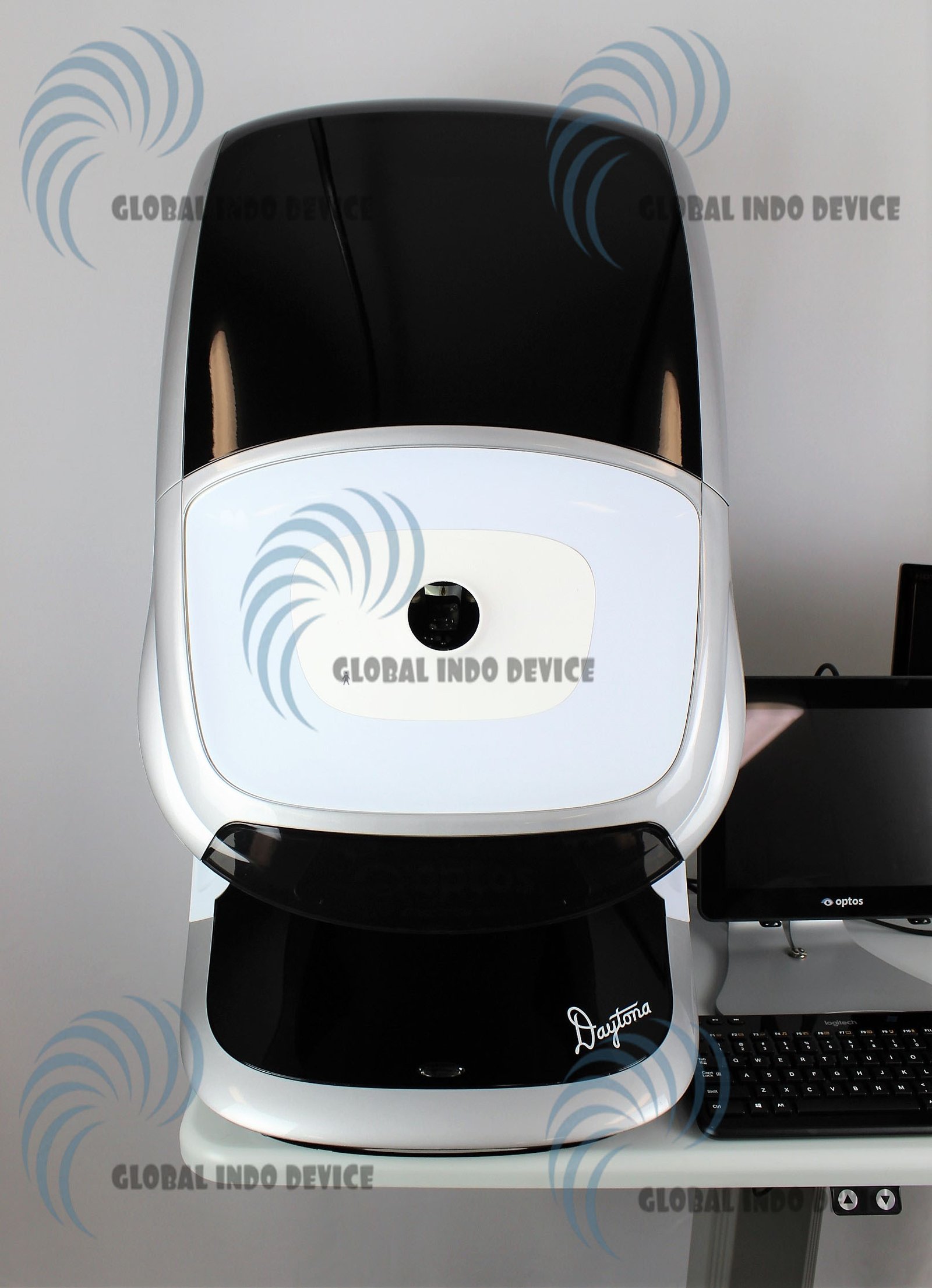
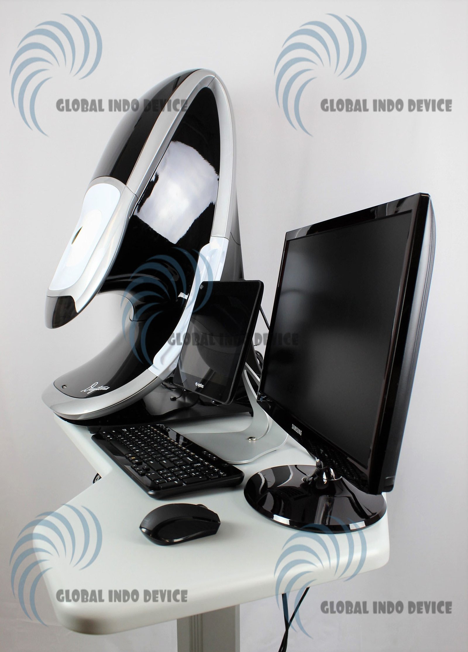
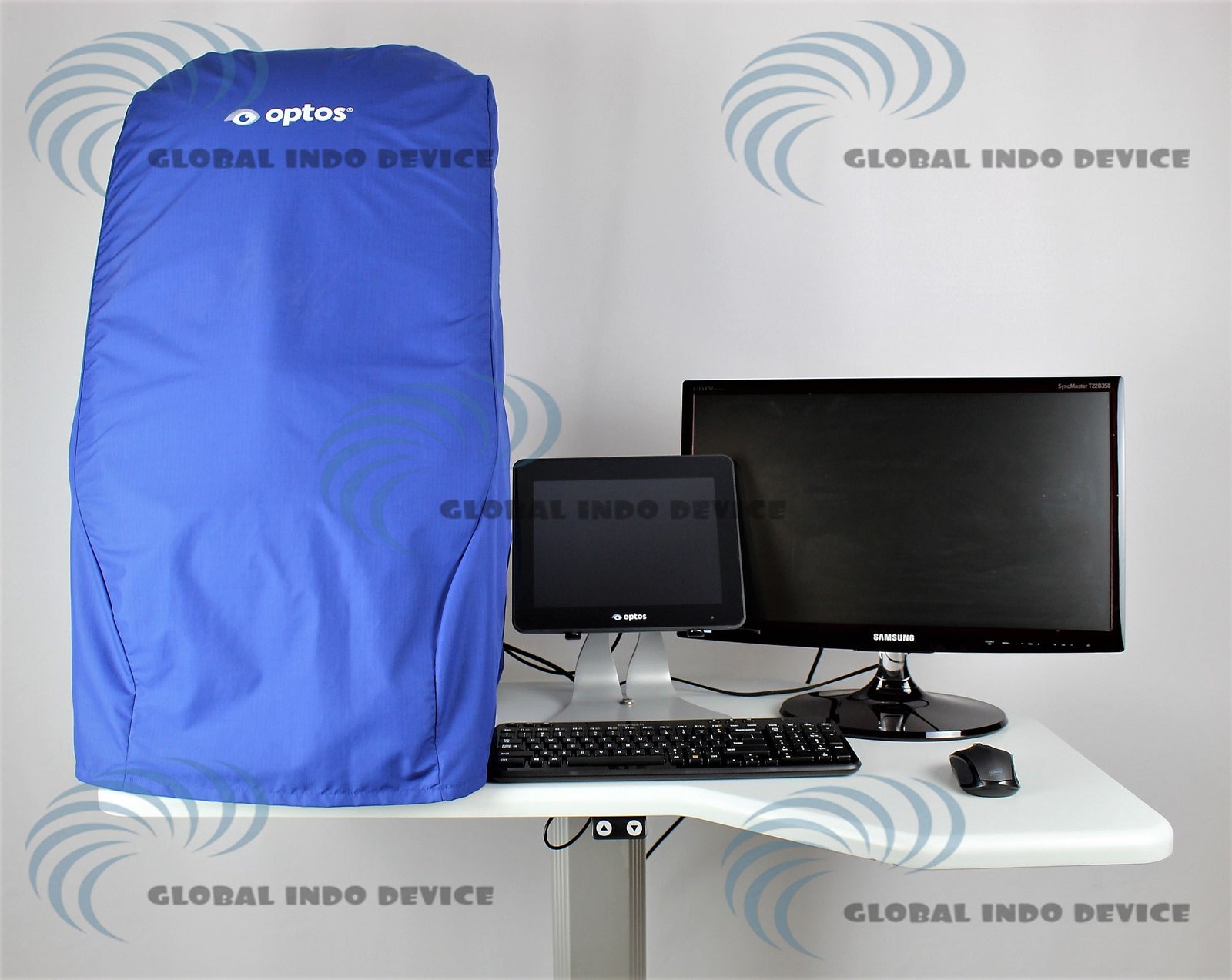
OPTOS Daytona Retinal Imaging Device
The OPTOS Daytona Retinal Imaging Device is an advanced ophthalmic imaging system designed to capture ultra-widefield retinal images quickly and efficiently. This technology supports early detection, diagnosis, and monitoring of retinal conditions while enhancing patient comfort and clinical workflow.
What Is the OPTOS Daytona Retinal Imaging Device
The OPTOS Daytona Retinal Imaging Device is a professional retinal imaging solution developed for eye care clinics and hospitals. It enables clinicians to obtain detailed retinal views without the need for pupil dilation in many cases.
Ultra-Widefield Retinal Imaging Technology
The device captures a wide view of the retina in a single image, allowing clinicians to visualize peripheral retinal areas that traditional imaging methods may miss.
Non-Mydriatic Imaging Capability
OPTOS Daytona often performs imaging without pharmacological dilation. This feature improves patient experience and reduces examination time.
Key Benefits of OPTOS Daytona Retinal Imaging
The OPTOS Daytona system provides clinical and operational advantages for eye care professionals.
Enhanced Retinal Visualization
Ultra-widefield imaging helps clinicians detect retinal abnormalities at earlier stages by providing a broader view of retinal health.
Improved Patient Comfort
Fast image capture and non-mydriatic operation reduce discomfort and anxiety for patients during retinal examinations.
Efficient Clinical Workflow
Quick image acquisition and easy operation allow clinics to increase patient throughput without compromising diagnostic quality.
How OPTOS Daytona Retinal Imaging Works
Understanding the imaging process highlights the efficiency of this system.
Scanning Laser Ophthalmoscopy
The device uses scanning laser technology to capture high-resolution retinal images. This method ensures consistent image quality with minimal light exposure.
Digital Image Processing
Captured images process digitally and integrate into electronic medical records, supporting documentation, comparison, and long-term patient monitoring.
Clinical Applications of OPTOS Daytona
The versatility of the system supports a wide range of ophthalmic examinations.
Retinal Disease Screening
Clinics use OPTOS Daytona to screen for diabetic retinopathy, retinal tears, and other peripheral retinal conditions.
Ongoing Retinal Monitoring
The system helps clinicians track disease progression and treatment outcomes through consistent imaging over time.
Ideal Users of the OPTOS Daytona Retinal Imaging Device
The system suits various eye care settings.
Ophthalmology and Optometry Clinics
Practices benefit from faster exams and improved diagnostic confidence with ultra-widefield imaging.
Hospitals and Vision Centers
High-volume facilities value the system’s speed, reliability, and ability to support comprehensive retinal evaluations.
Why Eye Care Professionals Choose OPTOS Daytona
Clinicians trust this imaging platform for its performance and diagnostic value.
Proven OPTOS Innovation
OPTOS is globally recognized for developing advanced retinal imaging technologies that support early detection and improved patient care.
High Diagnostic Confidence
Wide retinal visibility and consistent image quality help clinicians make informed clinical decisions.
OPTOS Daytona retinal imaging Device Features:
- Non-mydriatic, non-contact imaging through 2 mm pupils and many cataracts.
- High image resolution shows fine detail across the retina (optic disc, macula, and periphery).
- Autofluorescence imaging with green laser light displays lipofuscin in the RPE.
- Eyesteering further extends the field of view past the vortex vessels, in some cases.
- Stereo disc imaging.
- 3D Wrap for patient education
- DICOM compatible
- Innovative software tools enhance image evaluation.
- Images are available immediately and stored electronically for future comparison or for use in telehealth applications
- Image Modalities: Color, Red Free, Choroidal, Autofluoresence
OPTOS Daytona retinal imaging Device Benefits
– Improves Practice Efficiency and Economics: Studies show that optomap images are faster to capture and easier to review than traditional patient examination techniques 1, 2.
Optomap enables practitioners to differentiate their practice, and an additional revenue stream can be generated.
– Enhances Clinical Decision-making: Early signs of many ocular pathologies and diseases may first present in the retinal periphery and can go undetected using conventional techniques and equipment.
More than 400 published and ongoing clinical trials, thousands of case studies, and testimonials show the long-term value of optomap imaging in the diagnosis, treatment planning, and patient engagement.
− Helps Prevent Vision Loss through Technological Innovation: optomap technology can image pathology past the vortex vessels, helping practitioners find disease sooner and manage it more effectively.
Image views:
- Standard: 200° Single Capture
- Auto-montage: Up to 220⁰ (with OptosAdvance software only)
- Central Pole: Detailed view of the macula
- Stereo: Image pairing for optic disc and retinal evaluation
This Optos Daytona comes equipped with:
- Warranty through Optos after re-licensing machine
- Windows 10 OS with latest software
- Operators manual
- Power table
- Touchscreen tablet
There are no reviews yet.