
Product details
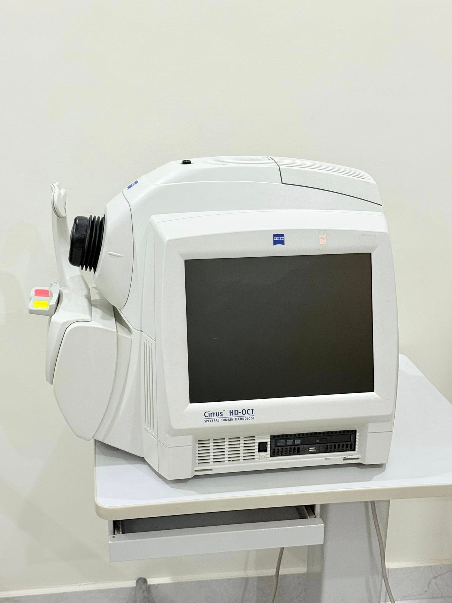
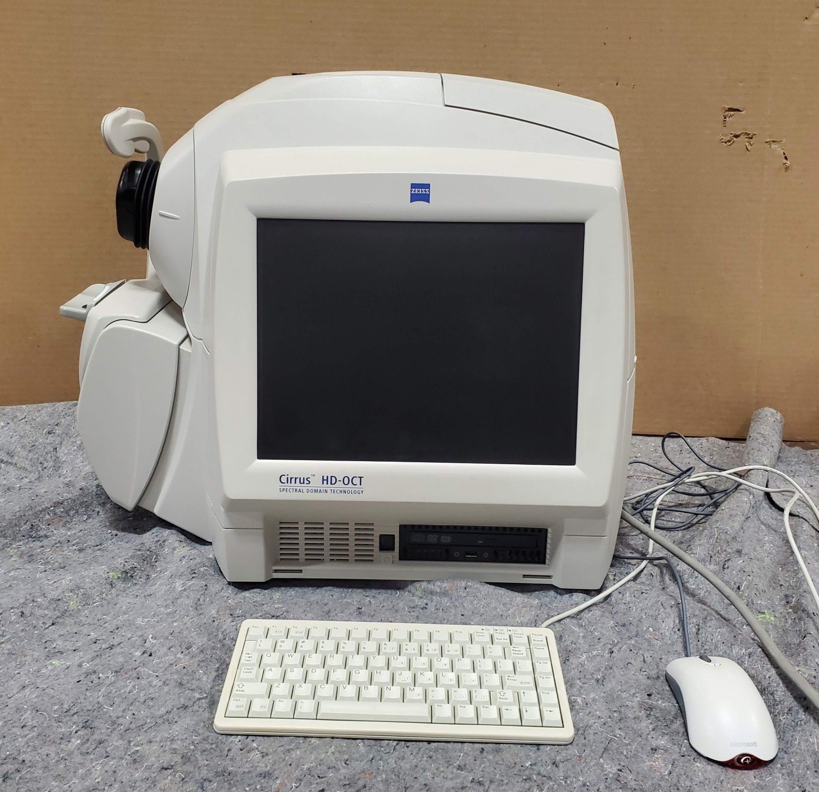
ZEISS CIRRUS 4000 HD-OCT: Advanced Retinal Imaging System for Precision Diagnostics
The ZEISS CIRRUS 4000 HD-OCT is a high-definition optical coherence tomography system designed to deliver precise, non-invasive imaging of the retina and optic nerve. Built for clinical efficiency and diagnostic confidence, this device combines advanced imaging technology with user-friendly operation, making it a reliable solution for ophthalmology practices and eye care clinics.
With its high-resolution scanning capabilities, the system enables clinicians to detect, monitor, and manage a wide range of ocular conditions, including glaucoma, macular degeneration, and diabetic retinopathy.
Key Features and Innovations
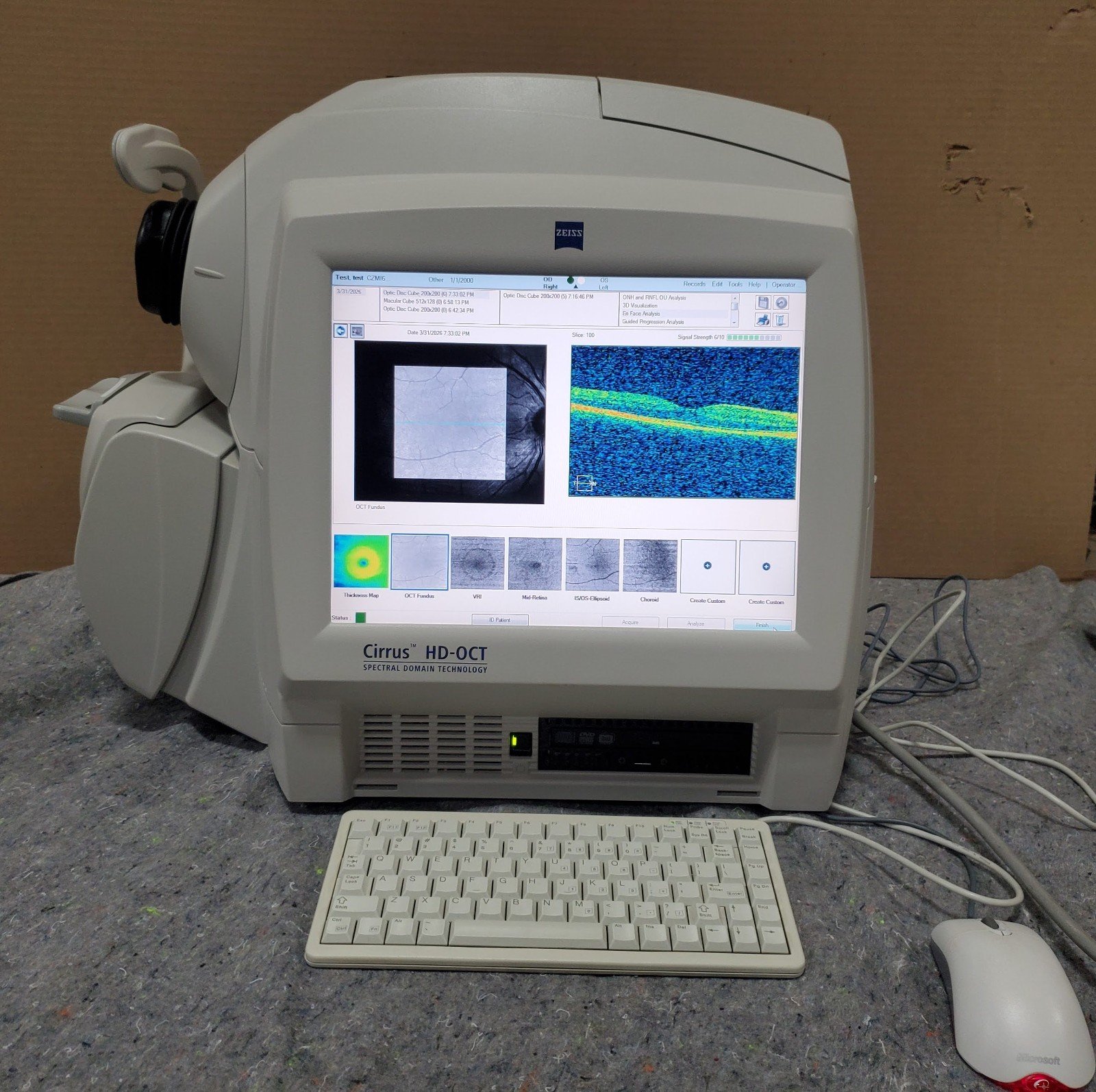
High-Definition Optical Coherence Tomography Imaging
The CIRRUS 4000 utilizes spectral-domain OCT technology to produce detailed cross-sectional images of retinal structures. This allows for early detection of subtle changes that may not be visible with traditional imaging methods.
Fast Scan Acquisition
The system offers rapid scanning speeds, reducing motion artifacts and improving patient comfort. Faster acquisition also increases workflow efficiency in busy clinical environments.
Advanced Analysis Software
Integrated software provides automated segmentation and measurement tools, enabling accurate assessment of retinal thickness, nerve fiber layer analysis, and macular mapping.
User-Friendly Interface
The intuitive interface simplifies operation, allowing clinicians and technicians to perform scans with minimal training. The system supports streamlined workflows and easy data management.
Reliable Data Management
Patient data is securely stored and can be easily accessed for comparison over time, supporting longitudinal studies and disease progression tracking.
Clinical Applications
Glaucoma Assessment
The device provides precise measurements of the retinal nerve fiber layer (RNFL), helping clinicians detect early signs of glaucoma and monitor disease progression.
Macular Disease Evaluation
High-resolution imaging allows detailed visualization of macular conditions such as age-related macular degeneration (AMD) and macular edema.
Diabetic Retinopathy Monitoring
The CIRRUS 4000 enables early detection of retinal changes associated with diabetes, facilitating timely intervention.
Optic Nerve Analysis
Detailed imaging of the optic nerve head supports accurate diagnosis and management of optic neuropathies.
Technical Specifications
Imaging Technology
- Type: Spectral-Domain Optical Coherence Tomography (SD-OCT)
- Light Source: Superluminescent diode (SLD)
- Central Wavelength: ~840 nm
Scan Performance
- Scan Speed: Up to 27,000 A-scans per second
- Axial Resolution: ~5 microns
- Transverse Resolution: ~15–20 microns
- Scan Depth: Approximately 2 mm in tissue
Scan Patterns
- Macular Cube Scan (512 × 128)
- Optic Disc Cube Scan (200 × 200)
- HD 5-Line Raster Scans
- Single Line Scan
- Crosshair Scan
Data Acquisition
- Acquisition Time: <2 seconds per scan
- Motion Artifact Reduction: Integrated tracking algorithms
- Signal Strength Indicator: Real-time quality feedback
Software and Analysis
- Automated Retinal Layer Segmentation
- Ganglion Cell Analysis (GCA)
- RNFL Thickness Mapping
- Optic Nerve Head Analysis
- Normative Database Comparison
- Progression Analysis Tools
Display and Interface
- Monitor: High-resolution LCD display
- User Interface: Graphical, touch and mouse-enabled
- Data Export: DICOM compatibility for integration with PACS systems
Connectivity
- USB Ports: Multiple for data transfer
- Ethernet: Network connectivity for data sharing
- Compatibility: Works with electronic medical record (EMR) systems
Patient Interface
- Chin Rest: Adjustable for patient comfort
- Fixation Target: Internal fixation system for accurate alignment
- Pupil Requirement: Typically ≥ 2.0 mm
Power Requirements
- Voltage: 100–240 V AC
- Frequency: 50/60 Hz
- Power Consumption: Approx. 300 VA
Physical Dimensions
- Weight: Approximately 30–35 kg (including table)
- Footprint: Compact design suitable for clinic environments
Advantages for Clinical Practice
Enhanced Diagnostic Confidence
The high-resolution imaging and advanced analytics provide clinicians with reliable data for accurate diagnosis.
Improved Workflow Efficiency
Fast scan speeds and automated analysis reduce examination time and increase patient throughput.
Longitudinal Patient Monitoring
The system allows comparison of historical data, making it easier to track disease progression and treatment outcomes.
Non-Invasive and Patient-Friendly
The imaging process is quick, painless, and does not require direct contact with the eye.
Why Choose ZEISS CIRRUS 4000 HD-OCT
The CIRRUS 4000 stands out as a dependable imaging solution for eye care professionals who require precision, efficiency, and ease of use. Its combination of advanced OCT technology, comprehensive analysis tools, and streamlined workflow makes it suitable for both small clinics and large ophthalmology centers.
Maintenance and Support
Routine Maintenance
- Regular calibration checks
- Cleaning of optical components
- Software updates for optimal performance
Technical Support
- Manufacturer support services
- Remote diagnostics capabilities
- On-site service options

There are no reviews yet.